Indian scientists create detailed atlas of the brainstem
Indian scientists mapped more than 200 brainstem clusters in 3D, using microscope images and eight markers to chart a region that keeps people alive.

Indian scientists at the Sudha Gopalakrishnan Brain Centre at the Indian Institute of Technology Madras have built a detailed three-dimensional atlas of the brainstem, one of the brain’s least understood regions. The map identifies more than 200 clusters of brain cells and nerve pathways, using high-resolution microscope images and eight chemical markers rather than costlier molecular techniques.
The brainstem occupies only a small part of the brain, but it controls functions that keep people alive. That makes the new atlas valuable well beyond anatomy: it gives researchers a reference point for studying how signals move through a region that has been difficult to chart with precision, and how that wiring may change in disease.
Its importance also lies in what it connects. Medical imaging shows the brain as a whole, while cellular pathology can reveal what is happening at the level of individual cells. The new atlas sits between those two views, offering a bridge that could help scientists compare scans with tissue-level changes more accurately. In a field where the best-known maps have often focused on the cortex or on broad brain networks, a detailed brainstem reference fills a gap that has long limited neuroscience.
India’s role in that work is becoming more visible. The National Brain Research Centre in Gurgaon is the country’s only institute dedicated to neuroscience research and education, and IIT Madras has already produced another major reference dataset, a cost-effective 3D fetal brain atlas built in 2024 from five fetal brains and more than 5,000 images. Together, those projects show that Indian labs are moving beyond data generation and into the creation of foundational research infrastructure.
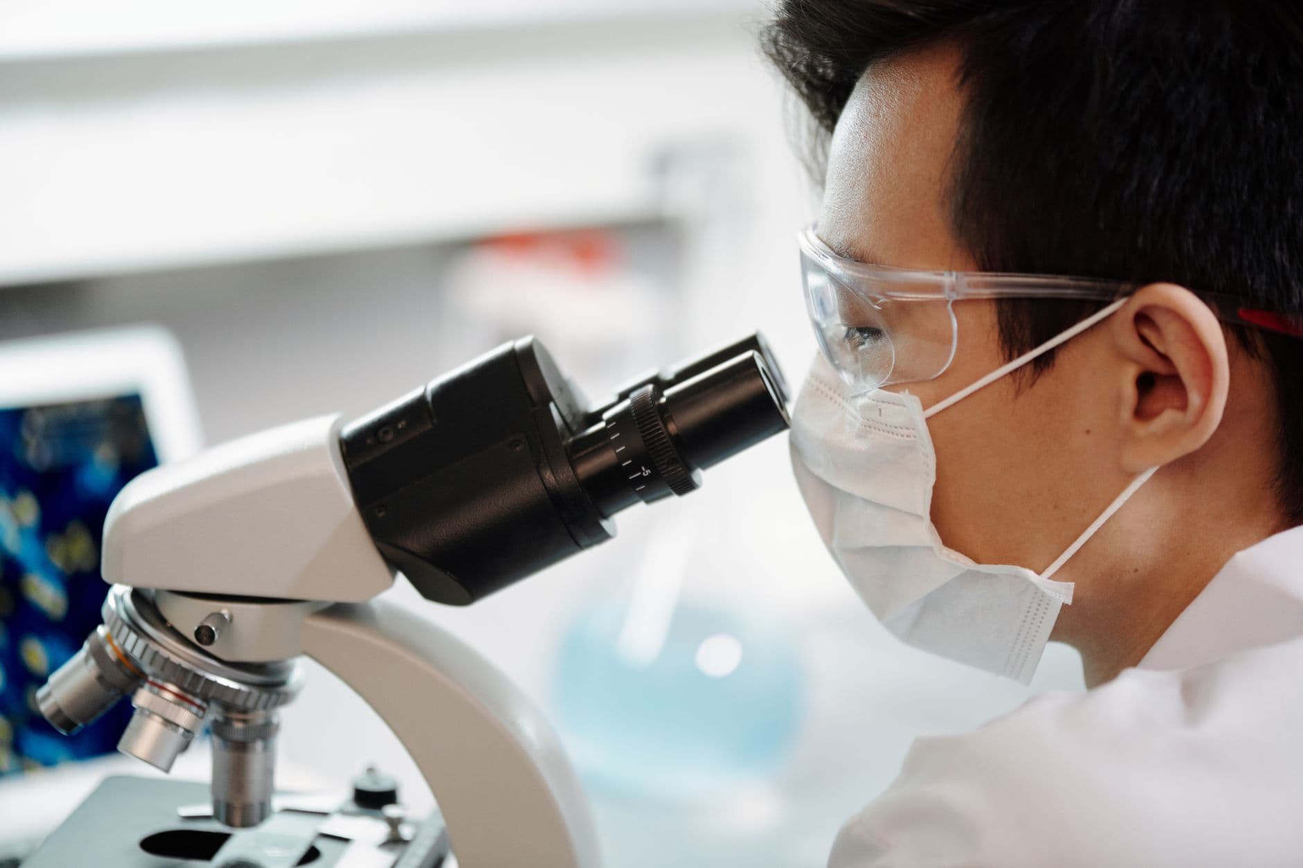
That shift comes as brain mapping accelerates worldwide. The Human Connectome Project began in 2010 with $40 million from the National Institutes of Health to map brain connections in young adults and later expanded across the lifespan. In 2023, researchers published the largest map of the human brain ever made, cataloguing more than 3,000 cell types. In 2024, Google researchers described a human brain tissue connectome containing about 16,000 neurons, 32,000 glia, 8,000 blood vessel cells and 150 million synapses in a sample about the size of half a grain of rice.
The new Indian atlas adds a highly specific resource to that race. It shows that some of the next major advances in neuroscience may come not only from bigger datasets, but from better maps of the brain’s least visible terrain.
This article was produced by Prism’s automated news system from verified source data, official records, and press releases, then run through automated quality and moderation checks before publishing. The system is built and supervised by the people who set the standards it runs under. Read our full AI policy.
Did this article answer your question?