Kelowna team prints patient-specific brachytherapy applicators with 3D printers
Physicians at B.C. Cancer Kelowna used 3D printing to produce MRI- and CT-based applicators for brachytherapy, improving dose accuracy and patient comfort.

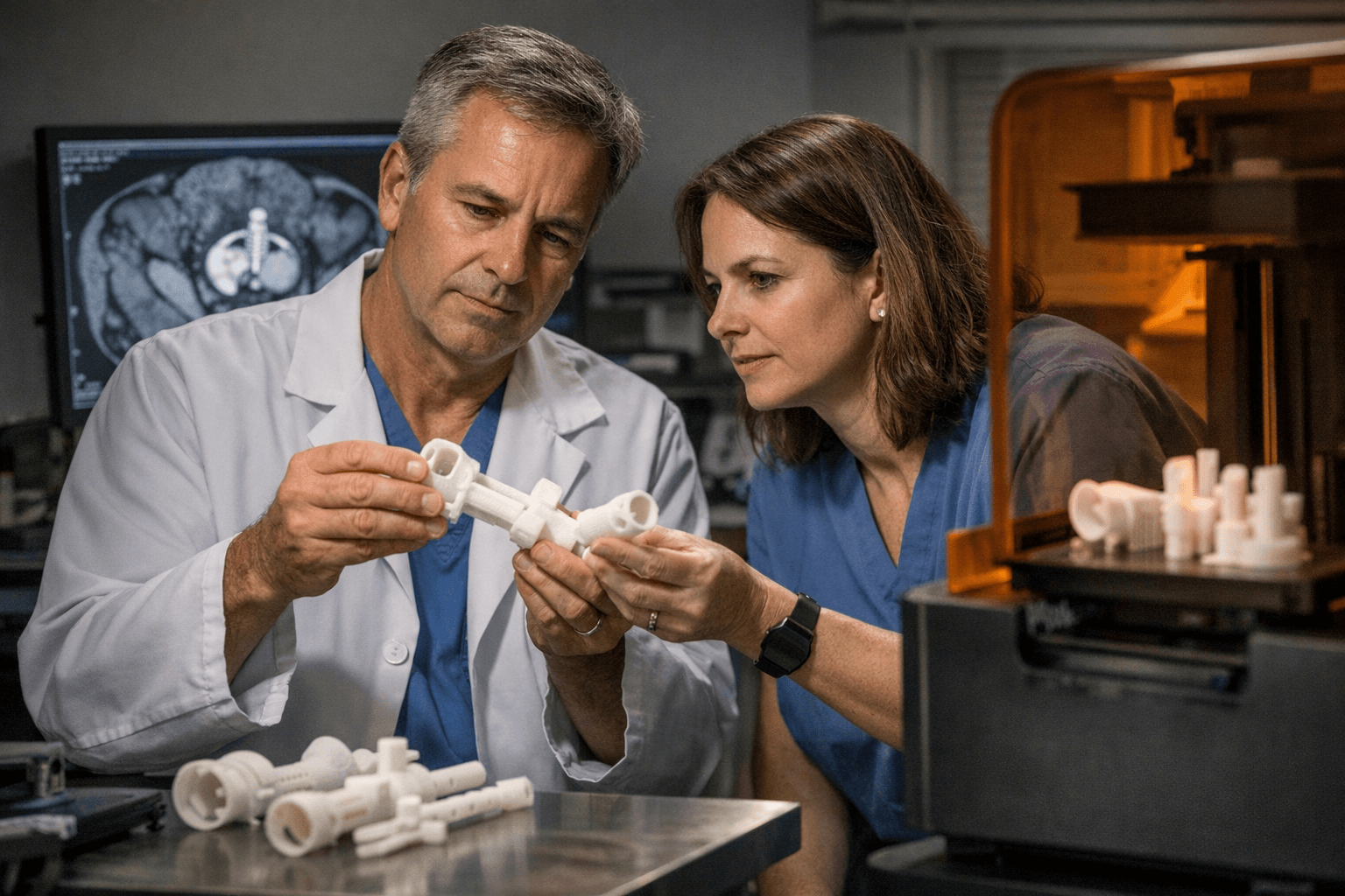
Physicians at B.C. Cancer Kelowna used a desktop 3D printer to create patient-specific applicators for brachytherapy, marking a first-in-Canada implementation of this imaging-to-print workflow. The custom devices, produced from each patient’s MRI and CT scans, let clinicians position radiation needles more accurately and deliver dose more precisely to cervical, vaginal and recurrent endometrial tumours while better protecting surrounding organs.
The project was led by medical physicist Dr. Deidre Batchelar, who also developed specialized software to convert imaging data into printable, patient-specific applicators. Clinicians at the Kelowna centre report that the bespoke applicators have already been used clinically with several patients and that the tailored fit reduces the number of needles required in some cases and improves patient comfort during procedures.
The 3D printer used in the centre cost approximately $60,000, a price point that makes this approach plausible for many regional hospitals and cancer centres with physics teams. By matching device geometry to each patient’s anatomy and tumour shape, clinicians say they gain tighter dosimetric control: higher effective dose to tumours with reduced exposure to organs at risk. That combination is expected to improve local control and decrease treatment side effects over time.
For the local maker and medical 3D printing communities, the Kelowna work underscores the practical value of desktop printing in clinical settings where customization matters. The workflow ties together standard cross-sectional imaging, in-house software development and desktop additive manufacturing to solve a very hands-on clinical problem: how to get needles to sit exactly where planned in complex pelvic anatomy. The result is not just a neat print but a procedural tool that changes how treatment is delivered on the table.
Adoption will depend on careful collaboration between radiation oncologists, medical physicists and institutional sterilization and regulatory processes, but the Kelowna example shows the baseline economics and technical pathway. A roughly $60,000 capital outlay and software that translates DICOM imaging into printable geometry can enable centres to test patient-specific applicators in routine brachytherapy streams.
For 3D printing practitioners and clinical teams, the immediate takeaway is clear: desktop printers are now crossing from prototyping into device-level clinical use where fit and dose matter. Expect more centres to pilot similar workflows, collect dosimetric and clinical outcomes, and refine software and printing protocols. If you’re in a hospital physics group, consider opening a conversation about imaging-based applicator design and whether a modestly priced printer could tighten your own brachytherapy plans and improve patient comfort.
This article was produced by Prism’s automated news system from verified source data, official records, and press releases, then run through automated quality and moderation checks before publishing. The system is built and supervised by the people who set the standards it runs under. Read our full AI policy.
Did this article answer your question?