Brain waste follows region-specific exit routes, study finds
Scientists traced a glowing protein out of mouse neurons and found the brain uses different exit routes by region, not one shared drain.

A fluorescent protein traced from mouse neurons has shown that brain waste does not leave through one universal drain. Instead, the new Cell study mapped region-specific exit routes through the dura, skull, nasal cavity and nearby lymph nodes, a pattern that points to a nearest-exit system rather than a single brain-wide pathway.
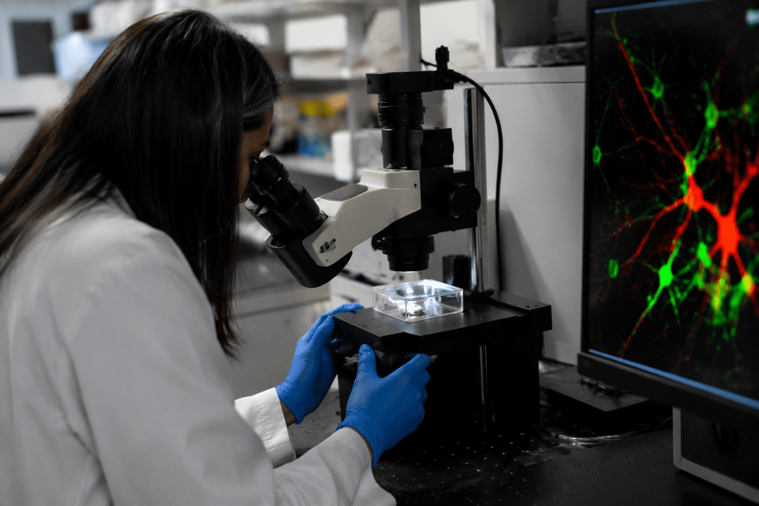
Andrew Yang of the Gladstone Institutes led the work, with Nalini Rao, PhD, and Yuichi Chayama, PhD, among the coauthors. Rather than inject tracers into cerebrospinal fluid, a method that can disturb the very movement scientists want to measure, the team engineered neurons in mice to make a fluorescent protein called ZsGreen and followed it as it left the brain. That switch matters because it let the researchers track protein produced inside the parenchyma itself, not just material moving through surrounding fluid.
The signal did not pour evenly toward one destination. Labeled protein moved into brain-adjacent borders, but very little reached the lymph nodes, undercutting the idea that most waste from the brain simply funnels to a single downstream sink. The picture that emerges is more local and more anatomical: different areas of the brain appear to send waste out through whichever border is closest, whether that is the dura, the skull or the nasal region. The study also reported that bordering immune cells interact with the waste products, and that Alzheimer’s disease disrupts this carefully organized clearance system.
That finding lands in the middle of a fast-moving shift in neuroscience. For years, researchers have argued over how the brain clears metabolic debris, especially proteins tied to Alzheimer’s pathology. Earlier NIH-supported work showed that parenchymal border macrophages help regulate cerebrospinal fluid flow and waste clearance in mice, and that removing those cells reduced flow and increased buildup of Alzheimer’s-related proteins. A 2024 study from Oregon Health & Science University added human data to the field, reinforcing that these clearance pathways matter beyond mouse models.
Maiken Nedergaard called the new paper a “game-changer,” saying it was the first study to directly detect endogenous protein efflux from the brain parenchyma rather than from cerebrospinal fluid. That distinction could reshape the next phase of the field, shifting attention from whether waste leaves the brain to how those pathways can be measured, predicted and possibly strengthened in living patients before toxic proteins accumulate.
This article was produced by Prism’s automated news system from verified source data, official records, and press releases, then run through automated quality and moderation checks before publishing. The system is built and supervised by the people who set the standards it runs under. Read our full AI policy.
Did this article answer your question?